Phasics
- Wavefront, MTF and QPI measurement solutions
- Products
- Applications
- Markets
- Company
- Contact us
Nov. 15, 2021
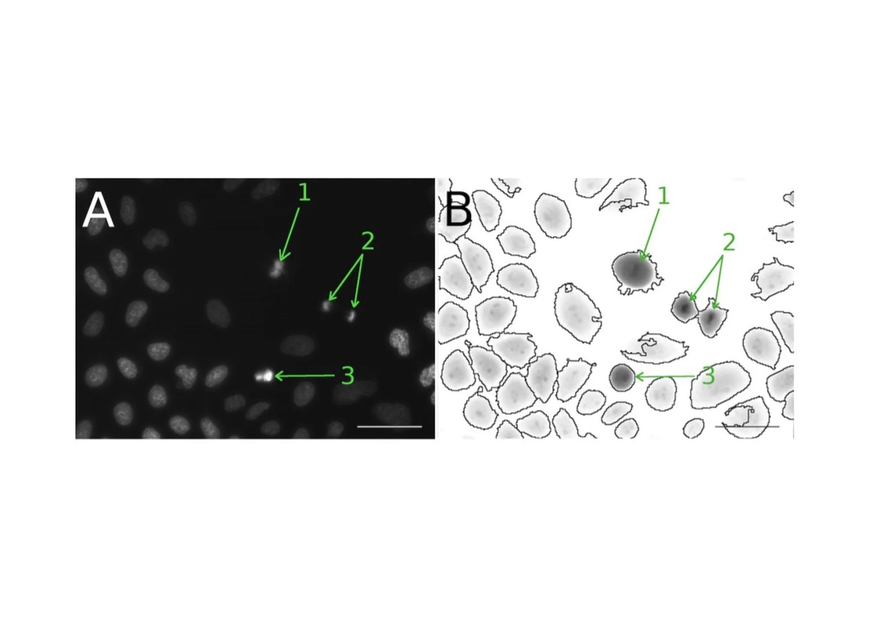
At the frontier of photonics, biology and artificial intelligence lies the future of life science. In a recent paper from the Université Côte d'Azur, researchers combine Quantitative Phase Images with a new supervised autoencoder to classify cells population based on the cell cycle. Images acquired with a Phasics SID4Bio QPI camera allows to increase the classification accuracy of cells present in the mitotic phase of the cell cycle.
Read the full article here: https://hal.archives-ouvertes.fr/hal-03364377
Learn more about PHASICS applications in life science here: https://www.phasics.com/en/application-markets/microscopy/life-science/
In the illustration (courtesy of Philippe Pognonec, Axel Gustovic, Zied Djabari, Thierry Pourcher, Michel Barlaud):